KPV (Lys-Pro-Val): Research Guide to the Anti-Inflammatory alpha-MSH Tripeptide
Dr. Sieglinde Klaus
Scientific Editorial Team · Bergdorf Bioscience

Table of Contents
- 01What is KPV and where does the tripeptide come from?
- 02How does KPV act at the molecular level?
- 03What antimicrobial activity is documented in studies?
- 04What dosages are used in research?
- 05How should the half-life and pharmacokinetics of KPV be assessed?
- 06How is KPV reconstituted and stored?
- 07What side effects are known from studies?
- 08How does KPV differ from BPC-157, Selank and Thymosin Alpha-1?
- 09Why does KPV not trigger pigmentation?
- 10What significance does PepT1 have for tissue selectivity?
- 11Frequently asked questions about KPV
- Is KPV approved for use in humans?
- Does KPV have a known half-life?
- Does KPV cause tanning of the skin?
- How should KPV be stored after reconstitution?
- How does KPV differ from Thymosin Alpha-1?
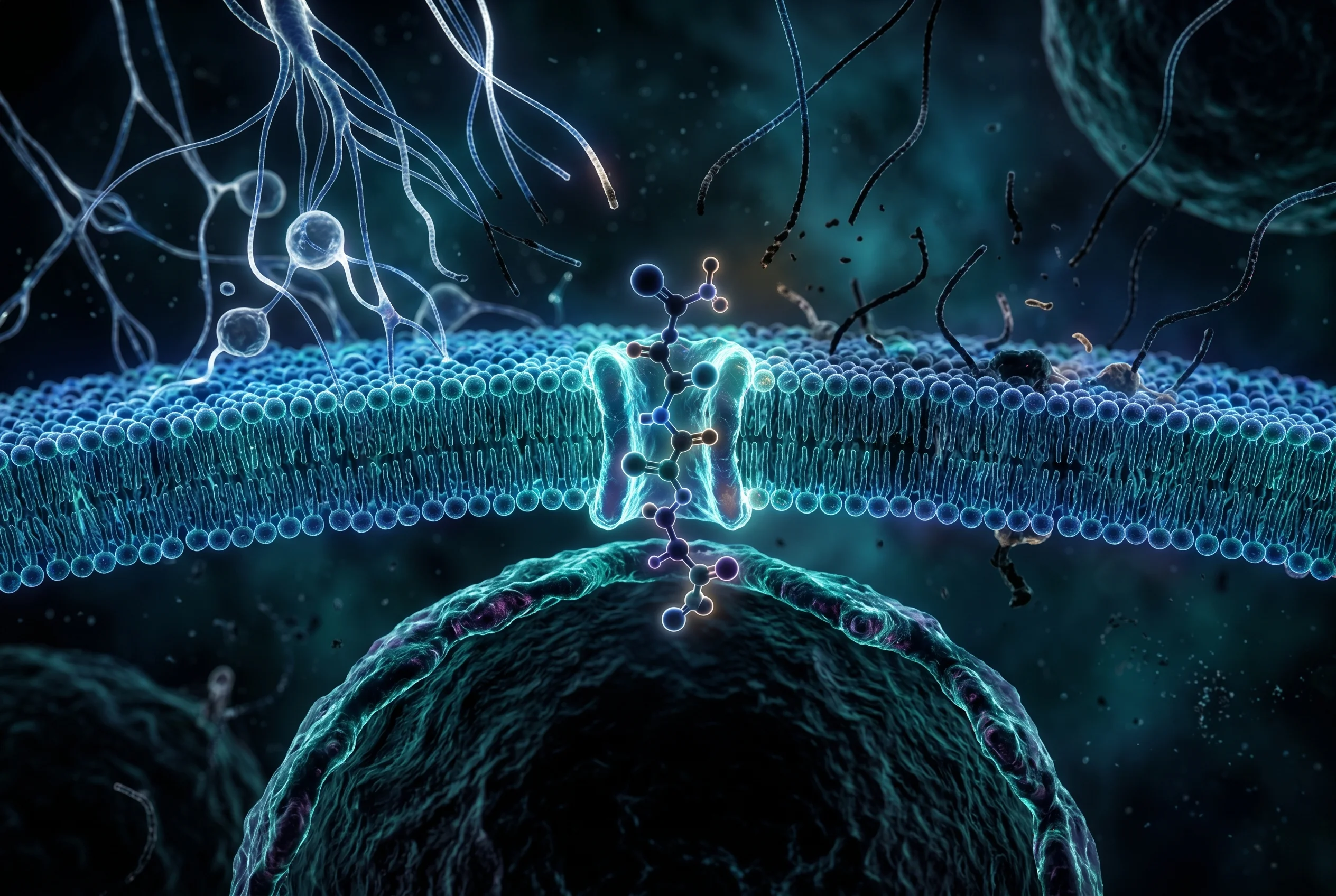
KPV is the C-terminal tripeptide (residues 11-13, Lys-Pro-Val) of alpha-melanocyte-stimulating hormone (alpha-MSH). Its defining property in research is a pronounced anti-inflammatory activity that is largely receptor-independent and runs intracellularly: the peptide is taken up into epithelial and immune cells via the di-/tripeptide transporter PepT1, where it blocks the nuclear translocation of the transcription factor NF-kB (Dalmasso et al., 2008).
What is KPV and where does the tripeptide come from?
KPV (single-letter code K-P-V) is a short, linear tripeptide made up of the three amino acids lysine, proline and valine. It corresponds to residues 11 to 13 at the C-terminus of alpha-melanocyte-stimulating hormone (alpha-MSH), a melanocortin. KPV therefore belongs to the class of melanocortin-derived peptide fragments. The molecular formula is C16H30N4O4 with a molar mass of approximately 342.44 g/mol for the free acid. Many research-grade preparations, however, are supplied in the N-acetylated and amidated form (Ac-KPV-NH2), which differs slightly in mass.
Notably, KPV carries the anti-inflammatory part of the alpha-MSH effect without triggering the pigmenting effects of the full hormone. Unlike alpha-MSH or Melanotan-type MC1R agonists, the tripeptide does not activate the cAMP-mediated MC1R response in melanocytes, so no pigmentation is initiated (Land, 2012). A distinct variant is the D-proline analogue Lys-D-Pro-Val (KdPV), which is protease-stabilized and used in some investigations (Haddad et al., 2001). In research practice, KPV is used above all as a model peptide for the intracellular modulation of inflammatory signaling pathways.
How does KPV act at the molecular level?
The dominant mechanism of KPV is intracellular and largely receptor-independent. The tripeptide is taken up into epithelial and immune cells via the human peptide transporter PepT1 (hPepT1). The affinity differs by cell type: in intestinal epithelial cells the Km value is around 160 uM, in Jurkat immune cells around 700 uM (Dalmasso et al., 2008). After uptake, KPV accumulates in the cell nucleus, where it competitively blocks the interaction between the NF-kB subunit p65/RelA and importin-alpha3 (at armadillo domains 7 to 8). This prevents the nuclear translocation of the NF-kB dimer (Land, 2012).
In addition, KPV stabilizes the inhibitor IkB-alpha by reversing its phosphorylation and degradation, and it inhibits MAPK signaling (Dalmasso et al., 2008; Haddad et al., 2001). On balance, nanomolar to micromolar concentrations suppress proinflammatory cytokines such as IL-6, IL-8, IL-12, IFN-gamma, IL-1beta as well as TNF-alpha-driven NF-kB reporter activity, without lowering the anti-inflammatory IL-10. A secondary, partly receptor-mediated component via MC3R/MC1R is described in airway and other epithelia (Dinparastisaleh and Mirsaeidi, 2021).
What antimicrobial activity is documented in studies?
Independently of the signaling effects described above, the C-terminal alpha-MSH sequence to which KPV belongs has a direct antimicrobial and antifungal activity. In investigations, this effect was observed against the bacterium Staphylococcus aureus as well as against the yeast Candida albicans (Cutuli et al., 2000). This property is mechanistically separate from NF-kB modulation and rests on the direct interaction of the peptide with the microorganisms.
In a broader framing, alpha-MSH and its derived fragments are discussed as an emerging class of anti-inflammatory and antimicrobial peptides that combine a dual function of immunomodulation and direct pathogen inhibition (Singh and Mukhopadhyay, 2014). This combination makes the tripeptide interesting for basic research, as it unites two otherwise separate principles of action in a very small molecule. The classification remains important: all of these findings come from in-vitro and model systems. There are no controlled human data that would support an antimicrobial application in humans. Studies merely point to a mechanistic potential that has been observed in preclinical systems and is the subject of further investigation.
What dosages are used in research?
For KPV there are no established therapeutic human doses; the peptide is not approved by the FDA. All reported research figures come from in-vitro and animal models. In cell culture studies, anti-inflammatory effects occurred across a broad concentration range of approximately 10 nM to 100 uM. In studies on airway epithelium, concentrations of 0.1 to 10 ug/mL were used, with a dose-dependent suppression observed from values of at least 1 ug/mL (Land, 2012).
In the mouse colitis animal model, KPV was administered at a concentration of 100 uM via the drinking water. This reduced DSS- and TNBS-induced inflammation, and myeloperoxidase activity as an inflammation marker fell by about 50 percent (Dalmasso et al., 2008). This local efficacy in the gut is explained directly by the PepT1-mediated cellular uptake at the tissue, and not by sustained systemic levels.
Vendor and clinic protocols circulate online, for example 200 to 500 mcg per day subcutaneously or oral 10-mg vials. These figures are not supported by published human PK or efficacy data and should be regarded as anecdotal and unvalidated. Anyone who wants to work with theoretical concentrations can follow them with KPV in the calculate KPV in the peptide calculator tool.

How should the half-life and pharmacokinetics of KPV be assessed?
An honest assessment is decisive here: in the peer-reviewed literature, KPV has no validated systemic half-life in humans. Plasma half-life, clearance, oral bioavailability and tissue distribution are practically uncharacterized in the human context. As an unprotected small peptide, KPV is rapidly hydrolyzed by peptidases. One study showed that acetylated KPV was completely broken down to its three amino acids within 24 hours by the enzyme pronase, which motivated the authors to a glycoalkylating modification to improve stability (Songok et al., 2018).
Frequently cited figures of about 30 minutes or roughly 0.5 hours of plasma half-life cannot be traced back to a primary human PK study. They appear to originate from secondary vendor pages and should be treated explicitly as unverified estimates and not as literature-backed values. Peptide agents of this size generically suffer from proteolytic instability and a short half-life. What is decisive for understanding is that the local efficacy of KPV, for example in the gut, is explained by the PepT1-mediated cellular uptake at the tissue and not by sustained systemic concentrations (Dalmasso et al., 2008). Anyone working with published values should document this uncertainty transparently.
How is KPV reconstituted and stored?
For KPV there is no peer-reviewed storage or temperature protocol. The following notes come from general handling recommendations for lyophilized peptides and not from primary literature. They are accordingly to be understood as precautionary measures and not as validated protocols. The lyophilized powder is typically kept at minus 20 degrees Celsius or colder for long-term storage. This recommendation follows the general handling of small peptides, whose known susceptibility to peptidases suggests protection against degradation.
After reconstitution with bacteriostatic or sterile water, the solution should be kept refrigerated at 2 to 8 degrees Celsius and used within a few weeks. Repeated freeze-thaw cycles are to be avoided, as is prolonged exposure at room temperature, since the proteolytic instability of the tripeptide is documented (Songok et al., 2018). When preparing, it is advisable to let the solvent run slowly down the wall of the vial rather than directing the stream straight onto the powder, in order to minimize mechanical stress. The purity of the starting material, usually stated at values of 98 percent or higher, should be documented via a certificate of analysis, since unregulated research suppliers can vary widely in quality and impurity profile. Clean documentation of every batch is indispensable for reproducible research results.
What side effects are known from studies?
For KPV there are no controlled human safety data. In review articles, alpha-MSH analogues are generally described as substances with a favorable safety profile, but a specific toxicology for KPV is not presented in detail (Dinparastisaleh and Mirsaeidi, 2021). An important distinguishing feature is that KPV, unlike the full alpha-MSH and unlike Melanotan-type MC1R agonists, does not trigger any pigmentation. This eliminates one of the frequently discussed properties of melanocortin-based peptides.
Reasonable theoretical and anecdotal concerns relate to reactions at the injection site, a possible immunogenicity, as well as the risk from product purity and impurities from unregulated research sources. Since KPV suppresses the transcription factor NF-kB and dampens innate cytokine responses, broad or chronic immunosuppressive effects are a plausible but as yet unstudied aspect. Studies suggest that the effect selectively affects proinflammatory signaling pathways, but long-term data on possible consequences of a sustained dampening of the innate immune response are entirely lacking. KPV is intended exclusively for research purposes and is not intended for therapeutic use in humans. Any work with the peptide should take place under laboratory conditions and with documented safety precautions.
How does KPV differ from BPC-157, Selank and Thymosin Alpha-1?
KPV is clearly set apart from other research peptides by its origin, size and mechanism of action. Compared with BPC-157 the difference is fundamental: BPC-157 is a synthetic fragment of 15 amino acids from the human gastric Body Protection Compound and is associated in research with angiogenesis via the VEGFR2-eNOS signaling pathway as well as with tissue, tendon and gut regeneration. KPV, by contrast, is a tripeptide of three amino acids characterized by a PepT1-dependent intracellular NF-kB blockade. Parent structure, size, transport route and primary mechanism differ entirely (Dalmasso et al., 2008; Land, 2012).
Selank is a synthetic heptapeptide analogue of the immunomodulatory peptide Tuftsin, developed as an anxiolytic and nootropic substance acting via GABAergic, monoaminergic and BDNF signaling pathways in the central nervous system. KPV is not neuroactive and acts on peripheral and epithelial NF-kB inflammation; origin and target system are unrelated. Thymosin Alpha-1, finally, is a thymic peptide of 28 amino acids that acts broadly immunostimulatory by promoting T-cell maturation as well as TLR and dendritic cell signaling. With its immunosuppressive, anti-inflammatory orientation, KPV stands at the opposite end of the immunomodulatory spectrum and differs entirely in size and source.
Why does KPV not trigger pigmentation?
This question is central to understanding the selectivity of KPV. The full alpha-MSH activates the melanocortin-1 receptor (MC1R) on melanocytes and thereby initiates a cAMP-mediated response that leads to melanin formation and thus to pigmentation. It is precisely this axis that Melanotan-type MC1R agonists exploit. KPV as a C-terminal tripeptide, however, carries the anti-inflammatory activity of the hormone without triggering the pigmentary MC1R-cAMP response in melanocytes (Land, 2012).
The reason lies in the dominant receptor-independent mode of action of the tripeptide. While pigmentation requires a full, classical receptor activation, the anti-inflammatory effect of KPV rests predominantly on the intracellular blockade of NF-kB nuclear translocation after PepT1 uptake. A secondary, partly receptor-mediated component via MC3R and MC1R is indeed described in airway and other epithelia, but it is not sufficient to start the pigmentary cAMP cascade in melanocytes (Dinparastisaleh and Mirsaeidi, 2021). For research, this separation means that anti-inflammatory properties of the alpha-MSH system can be studied without introducing the confounding pigmentary effect. This selectivity is one of the reasons why KPV has gained importance as a model peptide for melanocortin-mediated inflammation modulation.
What significance does PepT1 have for tissue selectivity?
The transporter PepT1 is the key to understanding why KPV can be locally effective despite a lack of systemic stability. PepT1 is a di- and tripeptide transporter expressed in intestinal epithelial cells and in immune cells. Through it, KPV enters the cell interior, where it exerts its anti-inflammatory effect. The affinity is tissue-dependent: in intestinal epithelial cells the Km value is around 160 uM, in Jurkat immune cells around 700 uM (Dalmasso et al., 2008).
This transporter-bound uptake explains the apparent paradox between rapid proteolytic instability and documented local efficacy. In the mouse colitis model, inflammation was reduced despite the known susceptibility of the peptide to peptidases, because PepT1 funnels the tripeptide directly into the cells at the affected gut tissue before systemic transport is even necessary. Efficacy is thus a question of local cellular uptake and not of systemic exposure. For research, this yields a clear mechanistic picture: tissues with high PepT1 expression, in particular the intestinal epithelium, are the most plausible model systems for the study of KPV. Studies suggest that the NF-kB blockade takes hold most efficiently where the transporter enables the intracellular accumulation of the peptide.
Frequently asked questions about KPV
Is KPV approved for use in humans?
No. KPV is not approved by the FDA or a comparable authority and has no established therapeutic human doses. All available data come from in-vitro and animal models, and the peptide is intended exclusively for research purposes (Dalmasso et al., 2008).
Does KPV have a known half-life?
A validated systemic half-life in humans does not exist in the scientific literature. Frequently cited values of about 30 minutes cannot be traced back to a primary human PK study and are considered unverified estimates. As an unprotected tripeptide, KPV is rapidly broken down by peptidases (Songok et al., 2018).
Does KPV cause tanning of the skin?
No. Unlike the full alpha-MSH or Melanotan-type MC1R agonists, KPV does not trigger the pigmentary MC1R-cAMP response in melanocytes and thus carries the anti-inflammatory activity without pigmentary effects (Land, 2012).
How should KPV be stored after reconstitution?
As a precautionary recommendation from general peptide handling, the reconstituted solution is kept refrigerated at 2 to 8 degrees Celsius and used within a few weeks. Repeated freeze-thaw cycles and prolonged room temperature are to be avoided because of the documented proteolytic instability (Songok et al., 2018).
How does KPV differ from Thymosin Alpha-1?
KPV acts anti-inflammatory and immunosuppressive by dampening NF-kB and proinflammatory cytokines. Thymosin Alpha-1, by contrast, is a considerably larger peptide of 28 amino acids and acts immunostimulatory. Both stand at opposite ends of the immunomodulatory spectrum (Dinparastisaleh and Mirsaeidi, 2021).
For research purposes only. Not intended for human consumption. Scientific editing: Dr. Sieglinde Klaus
References
- Dalmasso G., et al. PepT1-Mediated Tripeptide KPV Uptake Reduces Intestinal Inflammation. Gastroenterology. 2008.DOI
- HADDAD J., et al. α-Melanocyte-related tripeptide, Lys-d-Pro-Val, ameliorates endotoxin-induced nuclear factor κB translocation and activation: evidence for involvement of an interleukin-1β193–195 receptor antagonism in the alveolar epithelium. Biochemical Journal. 2001.DOI
- Land SC. Inhibition of cellular and systemic inflammation cues in human bronchial epithelial cells by melanocortin-related peptides: mechanism of KPV action and a role for MC3R agonists. International journal of physiology, pathophysiology and pharmacology. 2012.PMID
- Dinparastisaleh R., Mirsaeidi M.. Antifibrotic and Anti-Inflammatory Actions of α-Melanocytic Hormone: New Roles for an Old Player. Pharmaceuticals. 2021.DOI
- Cutuli M., et al. Antimicrobial effects of α-MSH peptides. Journal of Leukocyte Biology. 2000.