
VIP Peptide: Receptors, Signaling and Research
Dr. Sieglinde Klaus
Scientific Editorial Team · Bergdorf Bioscience
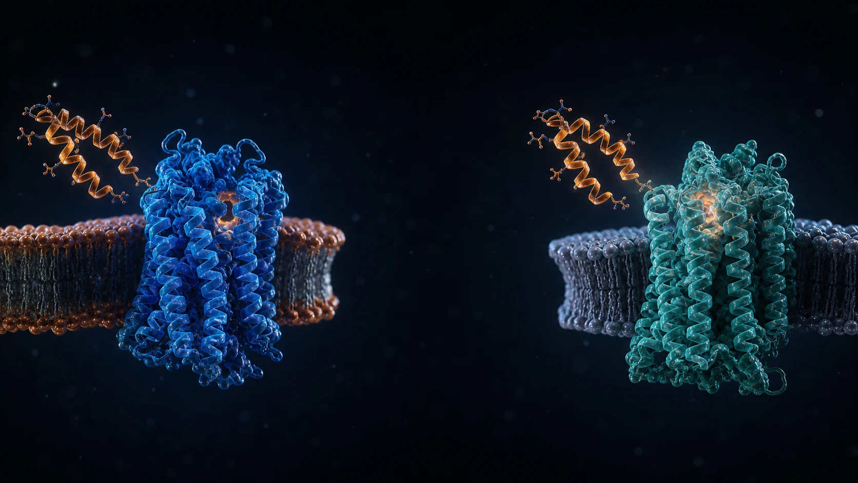
Table of Contents
- 01What is the VIP peptide and how is it structured?
- 02How does VIP recognize VPAC1 and VPAC2 receptors?
- 03How does VPAC activation lead to a cAMP signal?
- 04What does vascular research show about VIP?
- 05What does pulmonary VIP research find?
- 06What role is VIP research examining in the gastrointestinal system?
- 07Why are immunological statements about VIP especially context-dependent?
- 08What is known about VIP breakdown and pharmacokinetics?
- 09How can the state of VIP research be assessed responsibly?
- 10Which questions about the VIP peptide are frequently asked?
- Are VIP and vasoactive intestinal peptide the same thing?
- Which receptors does VIP primarily bind?
- Does cAMP activation mean the same VIP effect everywhere?
- Does VIP generally have a one-minute half-life?
- Do these studies establish a therapeutic use for VIP?
VIP, short for vasoactive intestinal peptide, is an endogenous 28-residue signaling peptide studied at the VPAC1 and VPAC2 receptors. In experimental systems, these receptors couple to Gs and cAMP signaling. What that signaling means depends on the receptor, cell type, tissue and model used, so these findings do not establish a therapeutic use.
What is the VIP peptide and how is it structured?
The name VIP stands for vasoactive intestinal peptide. The isolated peptide contains 28 amino-acid residues and has a C-terminal amide. In porcine-brain material, investigators determined the sequence HSDAVFTDNYTRLRKQMAVKKYLNSILN-NH2; the study also compared it with VIP previously isolated from upper intestine and with the amino-acid composition of human colon VIP (Carlquist et al., 1982). That sequence identifies the reference peptide; it does not establish the purity, folding, activity or biological effect of a particular research sample.
Biologically, VIP is produced from a larger precursor. Analysis of the human VIP gene identified seven exons, with the sequences for VIP and the related peptide PHM-27 located in separate exons of the same precursor gene (Linder et al., 1987). Precursor biology, the structure of the mature peptide and receptor interaction are therefore distinct research questions rather than interchangeable evidence.
The sequence alone does not predict a tissue outcome. In a systematic substitution study, individual residue changes affected binding and adenylyl-cyclase activation differently at human VPAC1 and VPAC2 (Nicole et al., 2000). These defined in-vitro receptor data show that particular residues can affect the measured assay; they do not predict organ-level activity.
For a research record, chemical characterization of the material under study should remain separate from biological reference literature. Isolation establishes primary structure, gene work addresses precursor organization, and receptor assays identify functionally relevant contacts in their stated system (Carlquist et al., 1982; Linder et al., 1987; Nicole et al., 2000). No one of these methods verifies every property of a separate sample.
How does VIP recognize VPAC1 and VPAC2 receptors?
VPAC1 and VPAC2 are class B1 G-protein-coupled receptors. Cloning of a human VIP receptor from HT29 cells showed specific, saturable VIP binding and a rise in cAMP after expression in COS-7 and 293 cells (Sreedharan et al., 1993). Characterization of the human VIPR2 gene described a seven-transmembrane receptor that was likewise coupled to cAMP production in functional systems (Lutz et al., 1999). These receptor entities are now termed VPAC1 and VPAC2.
Structural work adds molecular resolution without turning a receptor name into a complete effect description. Cryo-electron microscopy resolved an active, Gs-coupled VIP-VPAC1 complex, while complementary dynamics analyses reported mobile peptide contacts and distinctions from related PACAP27 complexes at VPAC1 (Piper et al., 2022). Ligand contacts, receptor conformation and the cellular background jointly shape the signal measured in an experiment.
VPAC1 and VPAC2 are not interchangeable labels. In an alanine-scanning study, each altered VIP residue was assessed for both ligand binding and adenylyl-cyclase activation at the two human receptor subtypes, revealing position-dependent differences (Nicole et al., 2000). Binding and activation are separate measurements: a substitution can affect affinity, signaling potency or both. A useful experimental account consequently identifies the ligand, receptor subtype, expression system and endpoint.
A cAMP response in a transfected cell line does not establish the same response in primary tissue, nor a clinical outcome. This boundary is fundamental to VIP research: molecular interaction is evidence at a molecular level. Tissue effects require their own functional evidence under their own conditions.
How does VPAC activation lead to a cAMP signal?
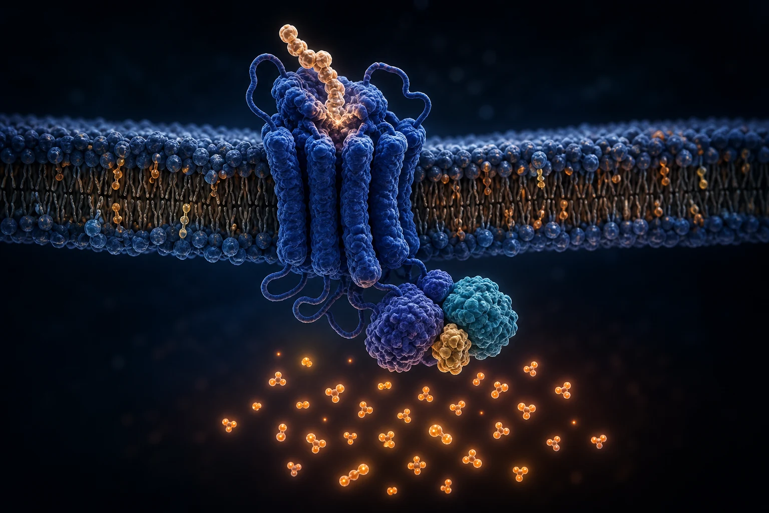
The best-supported early signaling route runs through Gs, adenylyl cyclase and the intracellular messenger cyclic adenosine monophosphate, or cAMP. Functional expression of human VPAC1 showed VIP binding together with a cAMP increase (Sreedharan et al., 1993). The active VIP-VPAC1 structure resolved in 2022 directly included Gs, linking functional observations to a structural snapshot (Piper et al., 2022).
cAMP is not, however, an organ-specific endpoint. In human Calu-3 bronchial epithelial cells, basolateral VPAC1 activation by VIP stimulated CFTR-dependent chloride secretion; inhibitor experiments placed PKA and PKC within the investigated pathway (Dérand et al., 2004). In a mouse intestinal immune-cell model, VIP on its own was weak, but with appropriate cytokine signals it potentiated selected ILC2 and ILC3 effector responses through cAMP and glycolytic energy mobilization (Pascal et al., 2022).
These findings explain why the statement “VIP raises cAMP” is mechanistically useful but biologically incomplete. Receptor density, compartmentalization, other signaling pathways and the cell's existing program influence the downstream result. Ligand binding, cAMP and the tissue-specific endpoint should therefore be measured separately. A single cAMP curve cannot stand in for vascular, pulmonary, intestinal or immune function (Dérand et al., 2004; Pascal et al., 2022).
Controls, pathway inhibitors, time course and concentration range help distinguish a VPAC-dependent response from other signals. Comparisons also require care: VIP-VPAC1, VPAC1-PACAP27 and PAC1-PACAP27 complexes differed in contact stability and dynamics (Piper et al., 2022). A VIP result at VPAC1 cannot simply be assigned to PACAP or PAC1.
What does vascular research show about VIP?
The word “vasoactive” reflects experimentally observed changes in vascular tone, not one uniform mechanism across all vascular beds. In a human skin study, VIP was examined locally by intradermal microdialysis. The observed vasodilation included a nitric-oxide-dependent component, and histamine H1-receptor blockade also reduced the response; H2 blockade did not have the corresponding effect in that model (Wilkins et al., 2004).
This evidence is local and method-specific. It concerns skin blood flow under microdialysis conditions and cannot be carried unaltered to coronary, cerebral or pulmonary vessels. Isolated-vessel work showed different dependencies: precontracted segments of human and bovine intrapulmonary arteries relaxed in response to VIP, and in that preparation the response did not require intact endothelium or adrenergic, cholinergic or cyclooxygenase-mediated signals (Greenberg et al., 1987).
The contrast marks a limit on comparability. Human skin microcirculation and isolated pulmonary-artery segments test different cell assemblies, starting-tone models and endpoints. The precise conclusion is that VIP was associated with relaxation under each experiment's conditions, while the implicated mediators depended on the vascular model. Neither study establishes a general blood-pressure effect or therapeutic suitability (Wilkins et al., 2004; Greenberg et al., 1987).
The measurements also differed. The skin study used laser-Doppler erythrocyte flux related to vascular conductance; the vessel preparation measured relaxation of precontracted artery segments. Although both endpoints are described as vasodilation, their scale and system are not the same. Interpretation should report the preparation, starting condition, measurement method and tested blockers.
What does pulmonary VIP research find?
Pulmonary VIP research addresses at least three distinct levels: pulmonary vessels, airway reactivity and epithelial function. In isolated human and bovine intrapulmonary arteries, VIP relaxed precontracted segments in a preparation where that response was largely endothelium-independent (Greenberg et al., 1987). This ex-vivo finding concerns vascular tone, not breathing or gas exchange in a living organism.
A genetic mouse model asked a different question. Mice lacking a functional VIP gene showed spontaneous methacholine airway hyperresponsiveness, peribronchiolar and perivascular cellular infiltrates, and elevated inflammatory markers in bronchoalveolar lavage (Szema et al., 2006). The finding supports a role for endogenous VIP signaling within that mouse model. A knockout changes a system throughout development, however, and is not equivalent to the short-term effect of externally supplied VIP in humans.
At the epithelial level, human Calu-3 bronchial cells expressed VPAC1. In Ussing-chamber and efflux experiments, VIP stimulated CFTR-dependent chloride transport when presented on the basolateral side (Dérand et al., 2004). The sidedness of the response and the cell-line system show why context must remain explicit.
Together, these studies are not a single chain of efficacy. They are separate observations in a vessel segment, a developmental mouse knockout and a human cell line. A vessel segment lacks complete neural regulation, ventilation and immune milieu; an epithelial cell line does not model a vascular response. Matching each claim to its model prevents “relaxation” and “inflammation” from being presented as one proven mechanism or a treatment claim.
What role is VIP research examining in the gastrointestinal system?
Gastrointestinal research extends beyond smooth-muscle relaxation. In over 2,200 surgical specimens from human small and large intestine, investigators studied neurally evoked epithelial secretion in vitro. Cholinergic contributions predominated in the small intestine, whereas VIPergic and nitrergic components shaped the response in the large intestine; transport involved cAMP-dependent chloride and bicarbonate secretion (Krueger et al., 2016). These were ex-vivo tissue experiments, not evidence about ingestion, treatment or a whole-body outcome.
Animal tissue also shows that receptor location and circuit arrangement matter. In guinea-pig jejunum, VPAC1 was involved in cholinergically mediated muscle-contraction responses and epithelial chloride secretion (Fung et al., 2014). That need not contradict a broad description of VIP as a relaxation-associated peptide: an excitatory interneuron can produce a different output from direct signaling in smooth-muscle cells. The preparation, cell type and endpoint resolve the apparent contradiction.
More recent mouse work adds a barrier-related neuroimmune circuit. Feeding-activated VIP-positive enteric neurons were positioned near VPAC2-expressing ILC3; in this model, VPAC2 signaling lowered IL-22 production in a defined ILC3 subset and changed downstream epithelial programs (Seillet et al., 2020). It is a feeding- and circadian-context mouse finding, not nutrition guidance or evidence of the same net effect in people.
Spatial resolution matters here. Human resection tissue distinguishes small from large intestine, the guinea-pig model concerns jejunum, and the ILC3 study defines immune-cell clusters in mouse ileum (Krueger et al., 2016; Fung et al., 2014; Seillet et al., 2020). “Gastrointestinal” alone is not a sufficient location when comparing secretion, motility or barrier findings.
Why are immunological statements about VIP especially context-dependent?
VIP cannot be described usefully in immunology as only suppressive or only activating. In a feeding-dependent mouse model, VPAC2 signaling reduced IL-22 production by a CCR6-positive intestinal ILC3 subset (Seillet et al., 2020). In another study, VIP alone weakly activated ILC2 and ILC3 but potentiated their response to additional cytokine signals through cAMP and glycolysis (Pascal et al., 2022). The receptor, subset, co-stimuli and endpoint are different.
A third mouse model added cell recruitment. VIP promoted intestinal ILC3 recruitment through VPAC1; mice lacking VIP or VPAC1 had fewer intestinal ILC3 and were more susceptible in a Citrobacter rodentium infection model (Yu et al., 2021). Recruitment through VPAC1 and cytokine regulation through VPAC2 answer different experimental questions. They do not amount to a general claim of immune enhancement or immune suppression.
The lung adds another context. In an allergic mouse-airway model, an IL-5-sensitive sensory-neuronal loop released VIP, stimulating CD4-positive cells and ILC2 and amplifying inflammation (Talbot et al., 2015). This does not make VIP generally pro-inflammatory, just as the inflammatory phenotype in VIP-knockout mice does not make it universally anti-inflammatory (Szema et al., 2006).
Organ, trigger, cell subset, receptor and time window must remain part of every statement. “Immunomodulatory” names a research field, not a predictable health effect. Cytokine amount, metabolic state and cell number are different endpoints. Keeping them separate makes the apparently mixed ILC findings coherent while avoiding conclusions that the underlying models did not measure.
What is known about VIP breakdown and pharmacokinetics?
One human observation came from a small study of four healthy volunteers. After graded intravenous VIP infusions ended, plasma VIP immunoreactivity declined with first-order kinetics. The authors reported a mean disappearance half-time of about one minute, apparent metabolic clearance of about 9 mL/kg/min and apparent volume of distribution of about 14 mL/kg (Domschke et al., 1978).
Every part of that statement carries context: four participants, intravenous infusion, a historical radioimmunoassay and a fall in measured plasma immunoreactivity after the infusion ended. “About one minute” is therefore not a universal systemic half-life. It does not describe another exposure route, tissue signaling, receptor binding or the duration of a biological effect. It also does not provide a dosing interval (Domschke et al., 1978).
The observation says nothing about a lyophilized material. Plasma disappearance is not vial shelf life, storage stability or stability after reconstitution. Formulation, matrix, temperature and analytical method belong to a stability study, but they were not product parameters in this human experiment. The one-minute result should not be turned into a general half-life-calculator input without a route qualification, or into a product description.
“Disappearance” is itself important wording. The study described the decline of VIP immunoreactivity measured by radioimmunoassay, not the direct loss of every intact peptide structure or receptor signal (Domschke et al., 1978). The measurement is assay- and route-bound. Transfer to tissue availability, other routes or duration of action would go beyond the design.
How can the state of VIP research be assessed responsibly?
Assessment starts at the evidence level. Sequence analysis defines the molecule; recombinant systems test binding and cAMP; cryo-EM describes receptor complexes; isolated tissues measure local functions; animal models connect responses across systems (Carlquist et al., 1982; Sreedharan et al., 1993; Piper et al., 2022). No level substitutes for another. Newer methods can add cellular or spatial detail without answering an earlier experiment's question.
Broad labels such as “vasodilatory,” “lung-protective” or “anti-inflammatory” need particular restraint. In human skin, VIP-associated vasodilation partly depended on nitric oxide and H1 signaling; isolated pulmonary arteries responded without an endothelial requirement in that preparation (Wilkins et al., 2004; Greenberg et al., 1987). In ILC models, cytokine signals could be reduced or potentiated depending on receptor, cell subset and co-stimulus (Seillet et al., 2020; Pascal et al., 2022).
Responsible VIP research identifies the species, tissue, receptor, experimental arrangement and direct endpoint. It separates endogenous VIP from externally investigated peptide. Bergdorf Bio research vials are not approved medicines and cannot be equated with them. For methodological context, see the guide to peptide side effects. This context does not convert these experiments into a recommendation for use.
Each claim remains tied to the material or genetic intervention, receptor subtype, species, tissue, immediate endpoint and the limits on extrapolation. For the human study, intravenous route, four participants and plasma immunoreactivity are indispensable qualifiers (Domschke et al., 1978). For cryo-EM, the purified receptor complex, Gs coupling and structural dynamics define the claim (Piper et al., 2022). Unmeasured levels remain open.
Which questions about the VIP peptide are frequently asked?
Are VIP and vasoactive intestinal peptide the same thing?
Yes. VIP abbreviates vasoactive intestinal peptide. The mature peptide has 28 amino-acid residues and a C-terminal amide (Carlquist et al., 1982). The abbreviation identifies the molecule, not a distinct receptor or signaling pathway.
Which receptors does VIP primarily bind?
VIP is investigated at VPAC1 and VPAC2. Recombinant human receptor systems show specific binding and cAMP coupling, while structural and substitution studies identify subtype differences in ligand recognition (Sreedharan et al., 1993; Nicole et al., 2000). The subtype must be demonstrated in each experiment.
Does cAMP activation mean the same VIP effect everywhere?
No. In bronchial epithelial cells, VPAC1-cAMP signaling was associated with CFTR-dependent chloride transport; in mouse intestinal ILC models, a defined response was potentiated only with additional cytokine signals (Dérand et al., 2004; Pascal et al., 2022). Cellular context determines the endpoint.
Does VIP generally have a one-minute half-life?
No. About one minute was the mean disappearance half-time of plasma immunoreactivity after intravenous infusions ended in four healthy volunteers (Domschke et al., 1978). It is neither route-independent nor a dosing interval or a statement about vial stability.
Do these studies establish a therapeutic use for VIP?
No. These studies address molecular, cellular, ex-vivo, animal or narrowly defined human research questions. Findings from VIP-knockout mice cannot be translated to externally supplied VIP in humans (Szema et al., 2006). Receptor binding alone does not establish clinical efficacy or safety.
For Research Use Only. Not for human consumption.
Scientific editing: Dr. Sieglinde Klaus
References
- https://pubmed.ncbi.nlm.nih.gov/7084607/
- https://pubmed.ncbi.nlm.nih.gov/36385145/
- Linder S, et al. Structure and expression of the gene encoding the vasoactive intestinal peptide precursor. Proceedings of the National Academy of Sciences of the United States of America. 1987.PMID
- https://pubmed.ncbi.nlm.nih.gov/10801840/
- https://pubmed.ncbi.nlm.nih.gov/8390245/
- Lutz EM, et al. Structure of the human VIPR2 gene for vasoactive intestinal peptide receptor type 2. FEBS letters. 1999.PMID
- https://pubmed.ncbi.nlm.nih.gov/14744818/