Subcutaneous Injection: Technique in the Research Lab
Dr. Sieglinde Klaus
Scientific Editorial Team · Bergdorf Bioscience
Dr. Sieglinde Klaus
Scientific Editorial Team · Bergdorf Bioscience
In a research context, subcutaneous injection refers to delivering a reconstituted solution into the subcutaneous fat layer of a model organism. In the lab this is done with fine insulin syringes (29G to 31G), under aseptic conditions, with a defined skin fold and systematic site rotation. This guide describes the technique strictly as handling of research material, not as instruction for human self-administration.
Subcutaneous (s.c.) refers to delivery into the loose connective and fatty tissue directly beneath the dermis, above the muscle fascia. Compared with muscle, this layer is poorly perfused, so substances pass into the capillary bed more slowly and more evenly. In a preclinical setting, the subcutaneous route is an established model for characterizing the absorption kinetics of peptides and proteins.
Absorption from the subcutaneous depot is slow and often incomplete: part of the substance is taken up via the lymphatic system, part is degraded in the interstitial space or by local enzymes before reaching the circulation (Richter & Jacobsen, 2014). This typically yields a lower peak concentration (Cmax) with a longer duration of action compared with intramuscular delivery.
Absorption rate depends strongly on the injection region. In clinical pharmacokinetic datasets, peptides with rapid absorption (Tmax less than or equal to 2 hours) and high clearance are especially sensitive to the choice of injection site (Zou et al., 2021). Reproducible research data therefore require a standardized, documented protocol. Before injection, the solution should be properly reconstituted; see the guide Reconstituting peptides for details.
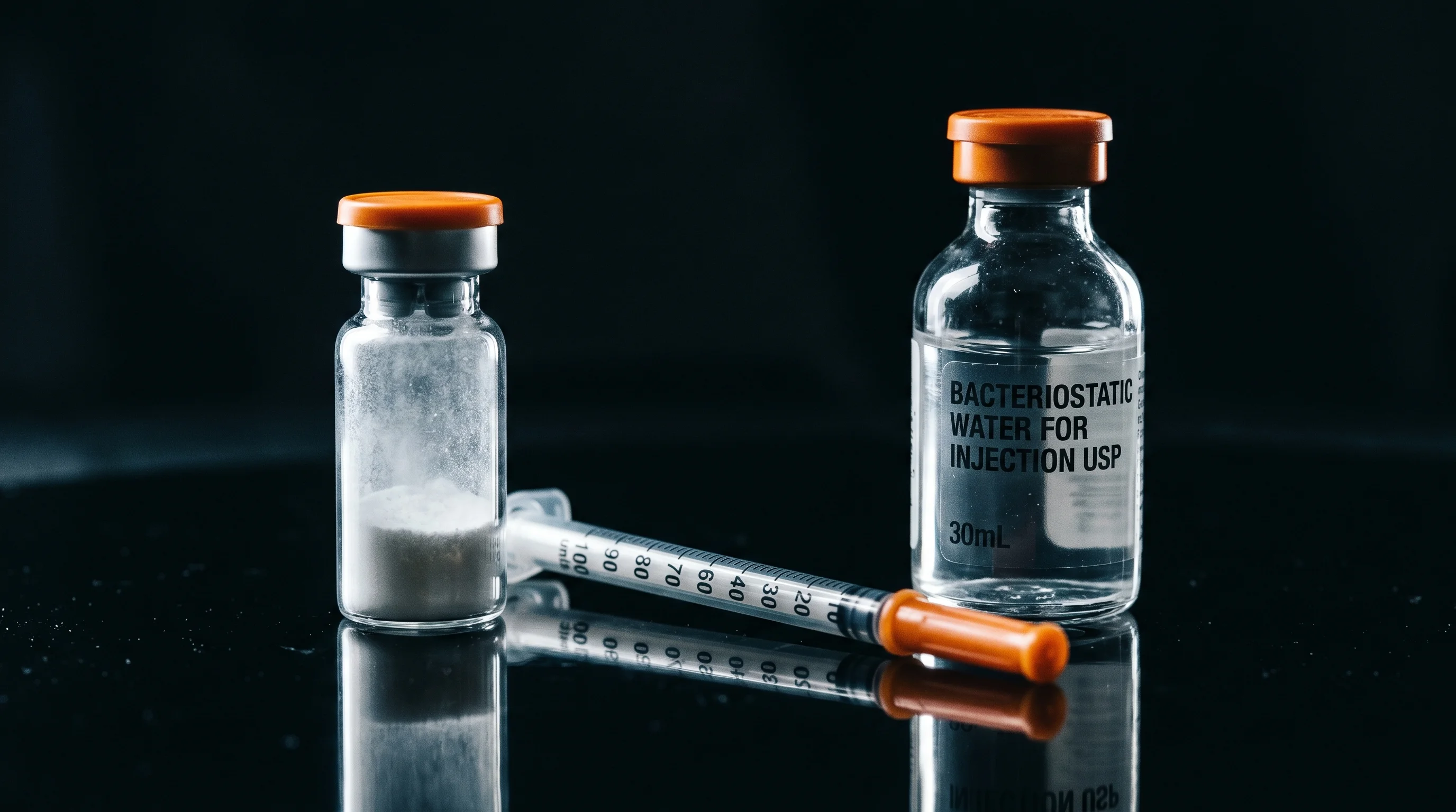
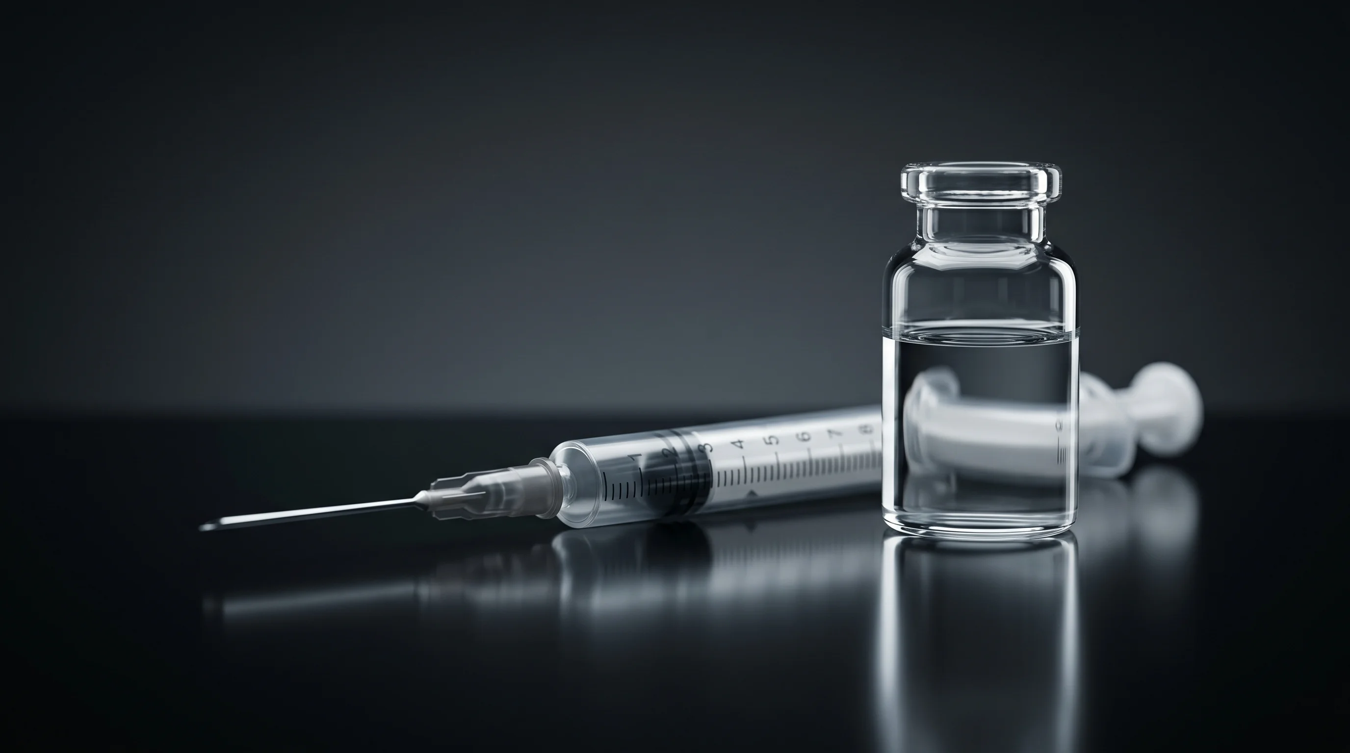
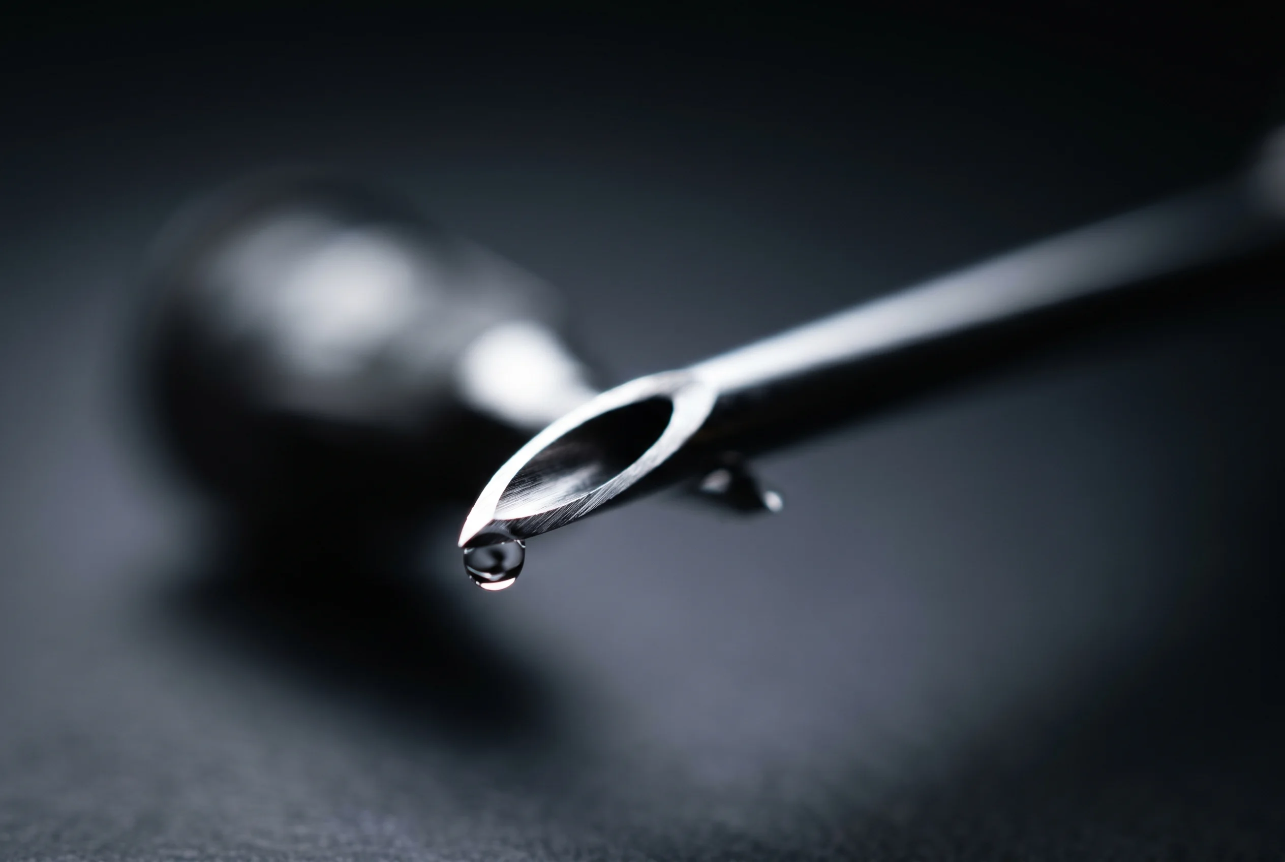
For subcutaneous applications in the lab, insulin syringes with a fixed integrated needle are standard. Needle thickness is given in gauge (G), where a higher number means a thinner outer diameter: 29G is about 0.33 mm, 30G about 0.30 mm and 31G about 0.25 mm. These fine needles create a small puncture channel and reduce tissue trauma and reflux.
Thinner needles are associated with less pain. In a controlled study of cutaneous needle insertion, the frequency of painful insertions rose significantly with outer diameter: 23G needles caused pain in 63 percent of insertions, 32G needles in only 31 percent (p less than 0.0001) (Arendt-Nielsen et al., 2006). This supports choosing the finest possible needles in animal models.
For drawing up: dose volumes exactly per calculation. The Peptide calculator helps convert concentration and target amount into units on the insulin scale. Air bubbles are removed by lightly tapping the barrel and gently expelling them before the needle reaches the skin fold. A separate, thicker draw-up needle can spare the fine s.c. needle, but with insulin syringes this is usually not part of routine practice.
The skin fold (pinch) lifts the subcutaneous tissue away from the underlying muscle and creates a defined depot. The skin is gently raised with thumb and index finger without grasping the muscle. With the fold raised, the needle is inserted at an angle of 45 to 90 degrees depending on needle length.
Needle length determines the angle: short needles (4 mm) can be inserted perpendicularly (90 degrees) with a skin fold raised, while longer needles often require a shallower angle of around 45 degrees to avoid piercing the muscle fascia. The FITTER recommendations identify the shortest needles (4 mm pen, 6 mm syringe) as safe and effective and stress that inadvertent intramuscular placement must be avoided, because it accelerates absorption kinetics and increases variability (Frid et al., 2016).
Penetration depth matters because tissue composition and depth influence uptake. In obese models, a needle that is too short may place the material too shallow, while one that is too long hits muscle (Erstad & Barletta, 2022). After insertion, the plunger is emptied evenly and completely; the skin fold is held throughout the injection and released only after the needle has dwelled briefly.
Repeated injection at the same spot alters the subcutaneous tissue and leads to lipohypertrophy, a thickening and hardening of the fat tissue. Absorption from such areas is blunted and highly variable, which distorts pharmacokinetic measurements and destroys comparability between experimental runs.
The evidence is clear: in a cohort of 372 subjects with type 1 diabetes, only 26.8 percent of those who rotated consistently developed lipohypertrophy, versus 83.9 percent without rotation; lack of rotation increased the risk 6.3-fold (Barola et al., 2018). The authors note that the blunted and variable absorption from lipohypertrophic sites leads to glycemic variability, a direct analog to data scatter in research.
In practice this means a documented rotation scheme across several distinct zones, with at least a few centimeters between successive insertions. Each used site is logged so that no area is loaded again before full recovery. This keeps depot formation and absorption conditions constant across the entire experimental series.
Beyond the injection site, several physiological and physical factors modulate uptake from the subcutaneous depot. A systematic overview names region, local blood flow, temperature, substance concentration and physical activity as the main determinants (Gradel et al., 2018).
Blood flow (subcutaneous blood flow) is central: increased flow recruits additional capillaries, enlarges the exchange surface and accelerates uptake. Temperature acts in the same direction. In the cited review, warming the injection site to 40 degrees Celsius reduced the time to maximum plasma concentration of insulin aspart by 42 percent. Physical activity of the model organism also raises regional perfusion and thereby the absorption rate.
Solution concentration behaves inversely: higher concentrations tend to slow uptake. Obesity delays absorption through lower capillary density. For reproducible research data these variables should be controlled, for example through constant ambient temperature, defined concentration and uniform regions. Storage of the solution also affects its integrity; see Storing peptides.
Aseptic work protects both the integrity of the research substance and the model against local reactions. Standard practice involves disinfecting the vial membrane and the skin site with a swab saturated with 60 to 70 percent alcohol, followed by adequate drying time. Only once the alcohol has fully evaporated is the puncture made, because wet alcohol in the puncture channel intensifies stinging and can irritate the substance.
The evidence on whether alcohol disinfection is strictly necessary before every subcutaneous injection is mixed; several reviews found no increased infection risk under clean conditions when alcohol was omitted. Nonetheless, aseptic work is considered good lab practice, especially with multi-dose vials. Each needle is used only once: reuse blunts the tip, increases tissue resistance and raises contamination risk.
Further basic rules: wear gloves, work on a disinfected surface, do not set down opened syringes and do not recap a needle to avoid sharps injuries. The membrane of multi-dose vials is wiped again before each withdrawal. These routines keep the microbial load low and ensure that observed effects are truly due to the substance and not to contamination.
The subcutaneous and intramuscular (i.m.) routes differ fundamentally in absorption profile and use case. Muscle tissue is far better perfused than the subcutaneous fat layer, so i.m. delivery of aqueous solutions typically reaches the circulation faster, produces higher peak levels and a shorter duration of action.
The subcutaneous route, by contrast, gives a slow, sustained profile with lower Cmax and a longer absorption phase, because the substance dwells in poorly vascularized fat and diffuses gradually (Richter & Jacobsen, 2014). For peptides and proteins this is often the preferred model route, since large molecules are additionally taken up via the lymphatic system.
Methodologically decisive: inadvertent i.m. placement when subcutaneous delivery was intended distorts data considerably because it accelerates onset. Precisely for this reason the FITTER recommendations stress short needles and the skin fold technique to reliably avoid the muscle fascia (Frid et al., 2016). In obese models needle length is especially critical, as misplacement is possible in both directions (Erstad & Barletta, 2022).
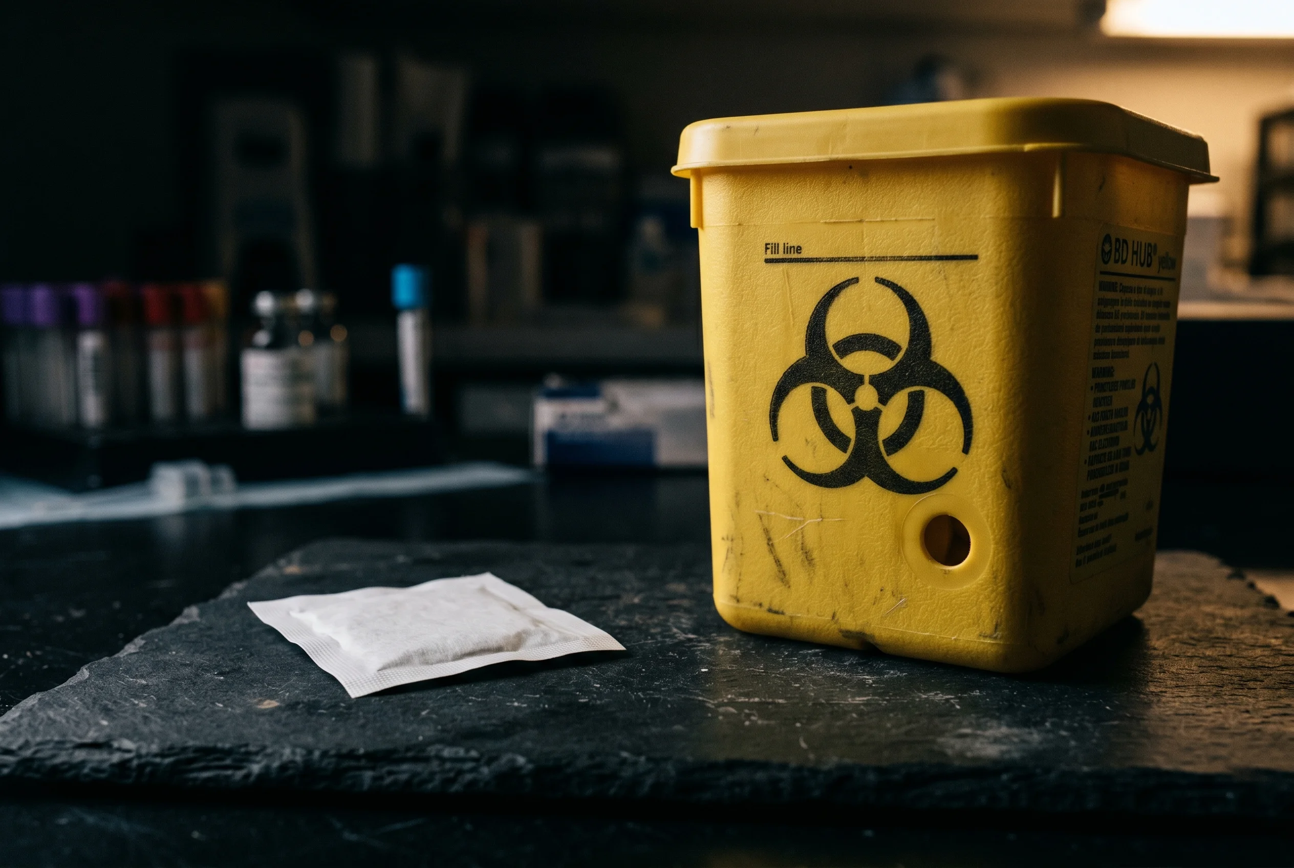
Used needles and syringes are sharp, contaminated objects (sharps) and belong without exception in a puncture-proof, sealable sharps container. This is placed directly at the workstation so the needle can be discarded immediately after use without an intermediate resting place.
The most important rule for avoiding needlestick injuries is: do not recap. Replacing the protective cap is one of the most common causes of accidental sharps injuries. The complete syringe including needle is discarded as a unit. The container is not filled beyond the fill line, because overfilled containers raise the injury risk on closing.
Disposal of the filled and sealed container follows the local biomedical waste regulations of the respective facility. Empty vials, swabs and packaging are separated according to laboratory waste guidelines. Complete documentation of batch, date and material used rounds off the protocol and ensures traceability for the research record.
A compact checklist secures reproducibility across all experimental runs. Before starting: solution properly reconstituted and checked for clarity, volume determined via the Peptide calculator, insulin syringe (29G to 31G) ready, sharps container within reach, work surface and gloves prepared.
During application: vial membrane and site disinfected and dried, air bubbles removed, skin fold raised, needle inserted at the appropriate angle (45 to 90 degrees depending on length), plunger emptied evenly, needle dwelled briefly, fold released. Site chosen per the rotation scheme and logged, with sufficient distance from the previous site.
After application: needle placed directly into the sharps container without recapping, material documented, work surface disinfected. Control variables such as temperature and concentration kept constant. This routine minimizes trauma, contamination and data scatter. It forms the methodological backbone for robust preclinical absorption data and complements the guides on Reconstituting peptides and Storing peptides.
For subcutaneous applications, insulin syringes of 29G to 31G are common. Higher gauge values mean thinner needles and are associated with less tissue trauma and less pain (Arendt-Nielsen et al., 2006).
The skin fold lifts the fat tissue away from the muscle and creates a defined subcutaneous depot. This prevents inadvertent intramuscular placement, which would accelerate absorption and increase data variability (Frid et al., 2016).
Each site should be reused only after full recovery. In one cohort, lack of rotation increased the lipohypertrophy risk 6.3-fold and leads to blunted, fluctuating absorption (Barola et al., 2018).
Aseptic work is considered good lab practice. The evidence on whether it is strictly required before every subcutaneous injection is mixed, yet disinfecting the vial and site with subsequent drying is recommended, especially with multi-dose vials.
Yes. Warming the injection site to 40 degrees Celsius reduced the time to maximum plasma concentration of insulin aspart by 42 percent (Gradel et al., 2018). Constant conditions are therefore important for reproducible data.
For research purposes only. Not intended for human consumption.
Scientific editor: Dr. Sieglinde Klaus