How to Reconstitute Peptides: A Step-by-Step Guide
Dr. Sieglinde Klaus
Scientific Editorial Team · Bergdorf Bioscience
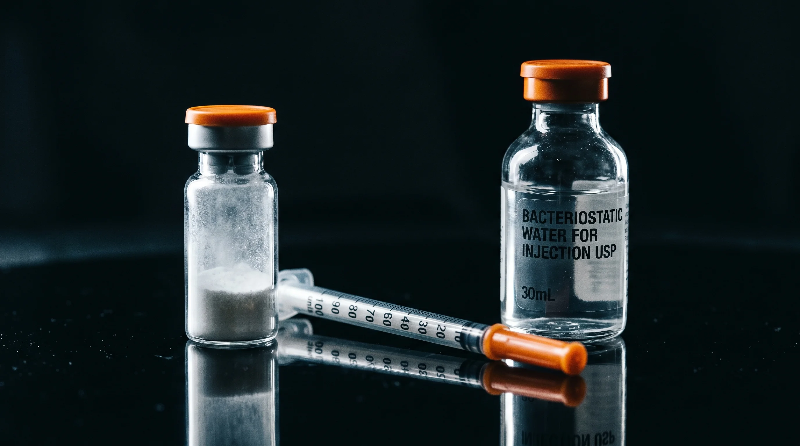
Table of Contents
- 01What does reconstitution mean for lyophilised peptides?
- 02What equipment do you need for reconstitution?
- 03How do you calculate the right reconstitution volume?
- 04How do you dissolve the peptide step by step?
- 05Why should you swirl instead of shake?
- 06How long does complete dissolution take?
- 07How do you store reconstituted peptides after mixing?
- 08What sterility rules apply during handling?
- 09What common mistakes occur during reconstitution?
- What water is suitable for reconstituting peptides?
- How do I calculate the right concentration?
- Why should you not shake the vial?
- How long is a reconstituted solution stable?
Reconstitution is the controlled process of dissolving a lyophilised (freeze-dried) research peptide in a suitable solvent, typically bacteriostatic water. At the bench, the dry substance is combined with a defined volume of liquid to produce a clear solution of known concentration (mg per ml). Clean, sterile technique is essential throughout to prevent contamination and degradation of the material.
What does reconstitution mean for lyophilised peptides?
Research peptides ship as a white, fluffy powder because freeze-drying removes water under vacuum, leaving behind an amorphous, storage-stable solid. In this dry state, the typical chemical degradation routes such as hydrolysis, deamidation and oxidation proceed far more slowly than in aqueous solution (Manning et al., 2010). Reconstitution returns this powder to a solution required for further laboratory handling. Once water is added, however, the chemical clock starts ticking: peptides in solution are inherently less stable than their lyophilised starting state. The goal of the procedure is therefore a reproducible, documented concentration combined with minimal microbial and mechanical stress. Typical amounts range from 1 to 10 mg of peptide per vial, dissolved in 1 to 5 ml of solvent. The chosen volume directly determines the final concentration and should be settled before the first handling step. Planning the target concentration in advance avoids later re-dilution, which adds extra pipetting steps and therefore extra sources of error. These steps are to be understood strictly as laboratory handling of research material.
What equipment do you need for reconstitution?
A short, complete materials list keeps the workflow uninterrupted. The following items belong on the bench:
- The vial of lyophilised peptide (usually 1 to 10 mg)
- A vial of bacteriostatic water as the solvent
- An insulin syringe with a fine needle, typically 29 to 31 gauge (G), graduated in units or ml
- Alcohol swabs (70 percent isopropanol) for disinfecting the rubber septa
- A clean, low-bioburden work surface, ideally inside a biosafety cabinet
- Optional: gloves and a small log sheet for batch, date and concentration
The fine 29 to 31 G needle reduces the size of the puncture in the rubber septum and therefore the risk of contamination and volume loss. Bacteriostatic water is the preferred solvent here because it contains 0.9 percent (9 mg per ml) benzyl alcohol as a bacteriostatic preservative, which inhibits microbial growth in the solution (FDA DailyMed, Bacteriostatic Water USP). If your supply runs out, you can order bacteriostatic water. If you are new to this class of substances, the primer What are peptides? offers context.
How do you calculate the right reconstitution volume?
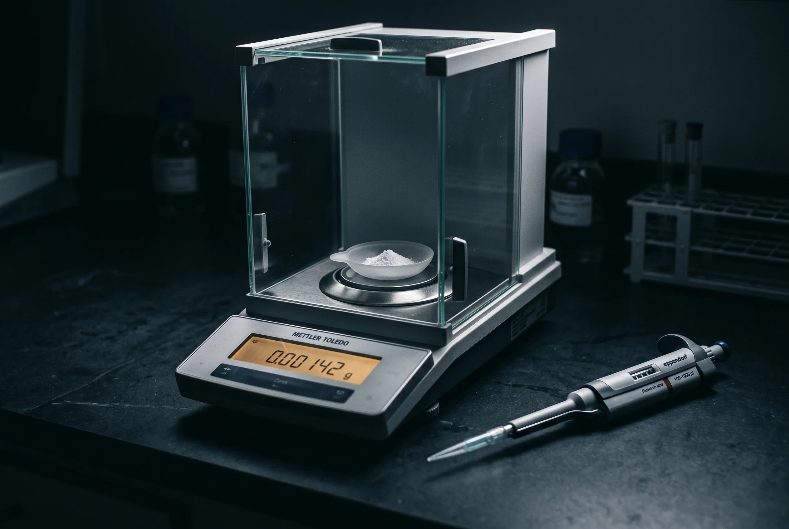
The core formula is straightforward: concentration equals the peptide mass divided by the added water volume, that is concentration (mg per ml) equals mg peptide divided by ml water. A vial of 5 mg dissolved in 2 ml of bacteriostatic water therefore yields 2.5 mg per ml. Add 5 ml instead, and the concentration drops to 1 mg per ml. The desired concentration depends on how finely you later need to measure volumes: a lower concentration means larger volumes to draw and therefore lower relative pipetting error. Because insulin syringes are often graduated in units (100 units equal 1 ml), it helps to choose a concentration so that the required amounts land on readable unit marks. A practical example: at 2 mg per ml, 10 units on the insulin syringe equal exactly 0.1 ml and therefore 0.2 mg of peptide. To avoid arithmetic mistakes and to model different volume scenarios, a digital aid is sensible. The peptide calculator handles the conversion between peptide mass, water volume and syringe graduation, reducing the risk of dilution errors. Write the calculated concentration directly on the vial so that every later withdrawal stays traceable.
How do you dissolve the peptide step by step?
The dissolution process follows a fixed order that protects both sterility and peptide integrity. Proceed as follows:
- Disinfect the rubber septa of both vials (peptide and water) with an alcohol swab and let them air-dry briefly.
- Draw the calculated volume of bacteriostatic water into the insulin syringe, for example 2 ml for a 5 mg vial.
- Pierce the needle through the peptide vial septum at an angle and aim the tip at the inner glass wall.
- Let the water run slowly down the vial wall rather than spraying it directly onto the powder. The stream must not blast the powder cake apart.
- Withdraw the needle and let the vial stand at room temperature until the powder dissolves.
Directing the stream onto the glass wall is not cosmetic; it reduces mechanical shear and the foaming that promotes aggregation at air-water interfaces (Zapadka et al., 2017). A forceful, direct jet of water can generate locally high shear forces and partially denature the peptide. Patience matters more than speed here. These instructions refer exclusively to the handling of research material at the bench.
Why should you swirl instead of shake?
After adding the water, undissolved material often remains at the bottom or on the wall of the vial. The temptation to shake vigorously is strong, but that is exactly counterproductive. Mechanical stress such as shaking, stirring or vortexing is an established tool in research for deliberately accelerating peptide and protein aggregation (Zapadka et al., 2017). Shaking creates countless small air bubbles and therefore a vastly enlarged air-water interface; this hydrophobic interface promotes the unfolding and clustering of peptide molecules. Instead of shaking, gently roll the vial between thumb and forefinger or swirl it in slow circular motions. For readily soluble peptides it is often enough to let the vial rest for a few minutes after adding the water; the powder then dissolves on its own. If a residue will not dissolve, repeated gentle swirling at intervals helps, but never aggressive shaking. A finished solution should be clear and free of visible particles, cloudiness or foam. If it appears turbid or forms flakes, this signals incipient aggregation or incomplete dissolution, and the sample should be evaluated critically. Gentle handling is therefore not a detail but a direct safeguard for molecular integrity.
How long does complete dissolution take?
Dissolution time depends strongly on the amino acid sequence, the peptide mass and the chosen concentration. Readily water-soluble, hydrophilic peptides often go fully into solution within a few minutes at room temperature, once the water has been added along the wall and the vial is gently swirled. Sequences with a high proportion of hydrophobic amino acids such as leucine, valine, phenylalanine or tryptophan dissolve more slowly and may need 15 to 30 minutes or repeated gentle swirling at intervals of a few minutes. The key is to give the process time rather than forcing it with mechanical violence. Accelerating dissolution by warming is not advisable, because elevated temperatures promote several degradation routes at once: deamidation and hydrolysis accelerate as temperature rises, and physical aggregation also shows a pronounced temperature dependence (Manning et al., 2010). If visible undissolved material remains after 30 minutes and several swirl intervals, the volume can be increased slightly to lower the concentration, or the sample is documented as incompletely dissolved. A fully reconstituted solution is optically clear; any persistent turbidity should be read as a warning sign. Build the dissolution time buffer into your experimental workflow from the start.
How do you store reconstituted peptides after mixing?
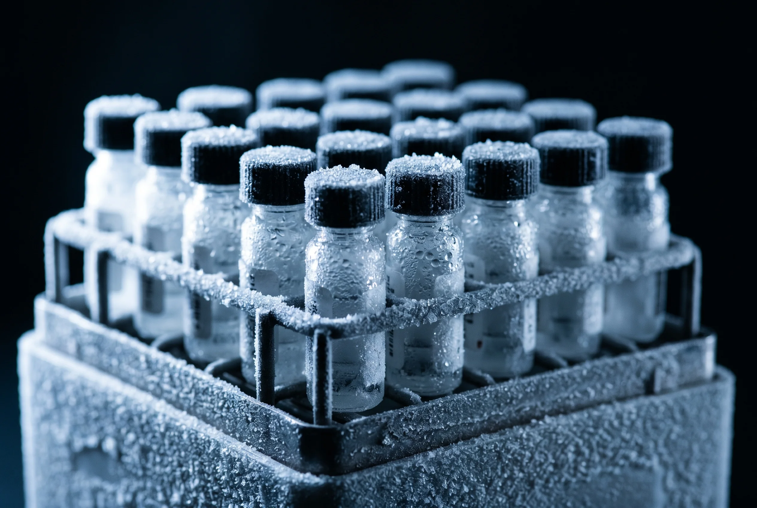
Once the peptide is in solution, its stability drops markedly, and storage conditions become the decisive factor. The reconstituted solution should be kept cool, dark and tightly sealed, usually in a refrigerator at 2 to 8 degrees Celsius. The benzyl alcohol in bacteriostatic water inhibits bacterial growth and thereby extends the usable window of the opened solution, but it does not replace clean technique (FDA DailyMed, Bacteriostatic Water USP). For longer-term storage, freezing is an option, but with one important caveat: every freeze-thaw cycle stresses the peptide mechanically and chemically. Studies on protein solutions show that each additional freeze-thaw cycle increases the particle count and therefore aggregate formation in the sample (Hauptmann et al., 2018). The practical consequence: divide the solution into small single-use aliquots before freezing, so that only one aliquot is thawed per use and repeated freezing is eliminated. Additionally protect the vials from light and label each with concentration and date. When thawing, slow warming in the refrigerator is preferable to a warm water bath, to avoid temperature shock. Following these rules keeps degradation low across the usable lifetime.
What sterility rules apply during handling?
Contamination control starts not at the moment of puncture but already with preparing the work surface. Clean the workspace, lay out all materials within reach and disinfect every rubber septum before each individual puncture with a fresh alcohol swab (70 percent isopropanol), allowing it to dry briefly. Use a sterile needle for each withdrawal and avoid passing the same needle repeatedly through different septa, as this can transfer microbes. Never touch the needle tip or the syringe cone with your fingers. The preservative benzyl alcohol does inhibit bacterial growth, but this protection is limited and not a licence for sloppy work; a bacteriostatic action does not kill existing microbes, it merely delays their multiplication (FDA DailyMed, Bacteriostatic Water USP). Note also that benzyl alcohol is not harmless: historically it was linked in premature infants to the so-called gasping syndrome, a severe toxic reaction from high benzyl alcohol exposure (Gershanik et al., 1982). This underlines that bacteriostatic water is a laboratory reagent with its own safety profile, used exclusively for handling research material. Document every withdrawal to keep sample integrity traceable across its entire usable life.
What common mistakes occur during reconstitution?
Many reconstitution problems trace back to a small set of recurring mistakes that are easy to avoid with a little attention. The most common are:
- Spraying water directly onto the powder cake instead of letting it run down the glass wall, which creates unnecessary shear.
- Vigorous shaking to speed dissolution, which promotes foam and an enlarged air-water interface and therefore aggregation (Zapadka et al., 2017).
- Incorrect volume calculation, so that the final concentration does not match the planned experimental design.
- Warming to accelerate, which promotes several degradation routes at once.
- Repeatedly freezing and thawing the same solution instead of dividing it into aliquots (Hauptmann et al., 2018).
- Missing labels, so that concentration, batch and date are no longer traceable.
- Failing to disinfect the septum before puncture, which raises contamination risk.
Working through these points systematically yields reproducibly clear solutions of documented concentration. A short checklist at the bench helps ensure no step is skipped, especially when several vials are processed in series. The combination of correct volume math, gentle handling and consistent sterility forms the foundation of all clean laboratory handling of research peptides.
What water is suitable for reconstituting peptides?
At the bench, bacteriostatic water is usually used because its 0.9 percent benzyl alcohol content inhibits microbial growth in the opened solution. Some hydrophobic or poorly soluble sequences may require different solvents. The choice should always follow the solubility profile of the specific peptide.
How do I calculate the right concentration?
Concentration equals mg peptide divided by ml water. A 5 mg vial with 2 ml of water yields 2.5 mg per ml. The peptide calculator handles this conversion along with the insulin syringe graduation and reduces arithmetic errors.
Why should you not shake the vial?
Shaking creates many air bubbles and enlarges the air-water interface where peptides aggregate more readily. Gentle rolling or swirling dissolves the powder just as well while protecting the molecular structure. Mechanical stress is even used deliberately in studies to accelerate aggregation.
How long is a reconstituted solution stable?
In solution, peptides are markedly less stable than as a powder. A refrigerated solution (2 to 8 degrees Celsius) should be used promptly; for longer storage it is frozen in single-use aliquots to avoid repeated freeze-thaw cycles. The exact profile depends on the sequence.
For research purposes only. Not intended for human consumption. Scientific editor: Dr. Sieglinde Klaus
References
- Gershanik J., et al. The Gasping Syndrome and Benzyl Alcohol Poisoning. New England Journal of Medicine. 1982.DOI
- Manning M., et al. Stability of Protein Pharmaceuticals: An Update. Pharmaceutical Research. 2010.DOI
- Zapadka K., et al. Factors affecting the physical stability (aggregation) of peptide therapeutics. Interface Focus. 2017.DOI
- Hauptmann A., et al. Impact of Buffer, Protein Concentration and Sucrose Addition on the Aggregation and Particle Formation during Freezing and Thawing. Pharmaceutical Research. 2018.DOI
- https://dailymed.nlm.nih.gov/dailymed/fda/fdaDrugXsl.cfm?setid=ccadcf46-6a6f-436b-9bbc-17e2983a335f
